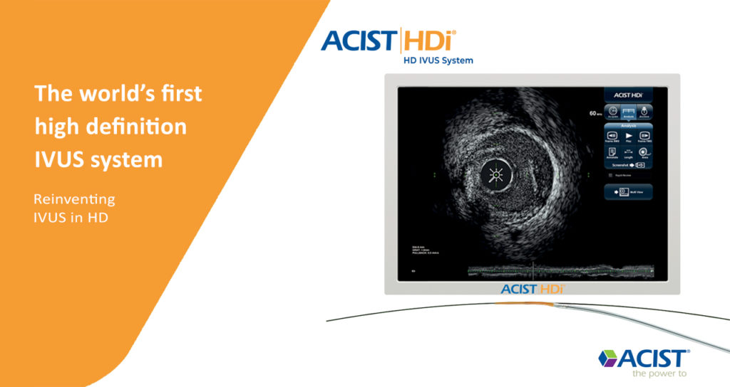
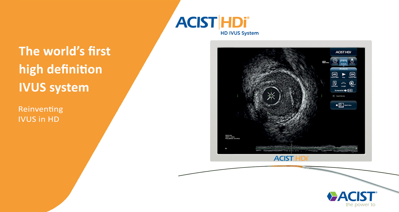
Intravascular ultrasound (IVUS) is a medical imaging methodology using a specially designed catheter with a miniaturized ultrasound probe attached to the distal end of the catheter. The proximal end of the catheter is attached to computerized ultrasound equipment. It allows the application of ultrasound technology, such as piezoelectric transducer or CMUT, to see from inside blood vessels out through the surrounding bloodcolumn, visualizing the endothelium (inner wall) of blood vessels in living individuals The arteries of the heart (the coronary arteries) are the most frequent imaging target for IVUS. IVUS is used in the coronary arteries to determine the amount of atheromatous plaque built up at any particular point in the epicardial coronary artery. Intravascular ultrasound provides a unique method to study the regression or progression of atherosclerotic lesions in vivo. The progressive accumulation of plaque within the artery wall over decades is the setup for vulnerable plaquewhich, in turn, leads to heart attack and stenosis (narrowing) of the artery (known as coronary artery lesions). IVUS is of use to determine both plaque volume within the wall of the artery and/or the degree of stenosis of the artery lumen. It can be especially useful in situations in which angiographic imaging is considered unreliable; such as for the lumen of ostial lesions or where angiographic images do not visualize lumen segments adequately, such as regions with multiple overlapping arterial segments. It is also used to assess the effects of treatments of stenosis such as with hydraulic angioplasty expansion of the artery, with or without stents, and the results of medical therapy over time. The ACIST HDi IVUS is a 60Mhz working frequency. Kodama Catheter has a frequency range of 40 and 60 MHz, specially designed for coronary and peripheral arteries. This kind of Catheter with various frequencies is unique to this company, which can meet all the physician's needs, including high image quality and a larger diameter display.
About HDi
IVUS reinvented in HD. High-definition 60 MHz IVUS imaging, touch panel interface, superfast pullback, and the highly deliverable ACIST Kodama HD IVUS Catheter.
Improved image quality
• Proprietary transducer and optimized signal processing for high-definition IVUS image quality, with minimized noise
• High-definition 60 MHz images of the vessel lumen and wall, without contrast flushing
• High depth of penetration to assess full plaque burden and the complete left main artery
Download Catalog

Benefits Of High-definition Ivus
Superfine axial resolution (<40 μm) versus other IVUS catheters (~100 μm) due to the 60 MHz transducer
Touchscreen enables rapid analysis
Up to 20× faster pullback reduces procedure time from minutes to seconds
Short distal tip, offset from the main catheter, improves lesion crossability and trackability and reduces the risk of guidewire entrapment and kinking


